
- Catalogs
- Hallmarq Veterinary Imaging
- Equine Referring Vet Brochure 2020

Equine Referring Vet Brochure 2020

Equine Referring Vet Brochure 2020
Catalog excerpts

REFERRING FOR EQUINE MRI Standing for Safety
Open the catalog to page 1
WHY ? WHAT’S SO SPECIAL ABOUT MRI? Since the advent of MRI, much has been learned about the causes of equine lameness. From the previously under-diagnosed, such as collateral desmitis of the distal interphalangeal joint, through the previously misunderstood, such as navicular syndrome, to the previously unknown, such as bone marrow edema, MRI has revolutionised our ability to provide a diagnosis and improve prognosis in equine lameness. and bruising in a way that has no parallel in radiography, CT, ultrasound or nuclear scintigraphy. Three dimensional imaging MRI images the region of interest...
Open the catalog to page 2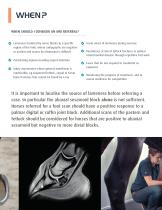
WHEN ? WHEN SHOULD I CONSIDER AN MRI REFERRAL? Lameness localised by nerve blocks to a specific region of the limb, where radiographs are negative or unclear and access by ultrasound is difficult Penetrating injuries needing urgent attention Injury assessment where general anesthesia is inadvisable, eg suspected fetlock, carpal or tarsal bone fractures that cannot be found by x-ray Acute onset of lameness during exercise Racehorses at risk of fetlock fractures or palmar osteochondral disease through repetitive fast work Cases that do not respond to treatment as expected Monitoring the progress...
Open the catalog to page 3
HOW ? HOW DO I REFER A CASE? The referral clinic will need to know the case history and previous diagnostic results. After the scan they will provide an interpretation and radiological report. The shoes are removed and, for foot scans, checks made for residual nail fragments The horse is sedated and stood in an electrically screened room with its leg inside a large magnet Scanning takes 1-2 hours, possibly longer if a horse is uncooperative. Then the horse will need some time to recover from sedation before going home Typically 500-600 images are collected, and interpreted by a specialist which...
Open the catalog to page 4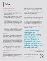
Q&A Are all MRI scanners the same? There are three types of MRI scanner: • Standing MRI. The Hallmarq system is the only MRI scanner that can image the foot and leg of the standing sedated horse. • Adapted human 1.5T high field, tubular scanners. Mostly found in institutions that scan both companion animals and horses. The reported diagnostic rate and lesions detected are similar for both high field and standing systems, though some clinicians prefer a high field machine for performance issues where only minor lesions are suspected. General anesthesia is always required. • Low field “down” scanners....
Open the catalog to page 5
“Standing MRI allows us to accurately diagnose the cause of lameness in the vast majority of cases where standard diagnostic techniques fail to give us the answer. It permits the selection of appropriate treatment methods, whereas without it we would often have been guessing” Tim Mair BVSc PhD DEIM DESTS DipECEIM MRCVS, Bell Equine Veterinary Hospital Hallmarq Veterinary Imaging, Inc. 1275 W. Roosevelt Rd., Suite 116 West Chicago, IL 60185, USA +1 978.266.1219 Hallmarq Veterinary Imaging Ltd Unit 5, Bridge Park, Guildford, Surrey, GU4 7BF, UK +44 (0)1483 877812 © Hallmarq Veterinary Imaging 2018....
Open the catalog to page 6All Hallmarq Veterinary Imaging catalogs and technical brochures
 2018 STANDING EQUINE MRI
8 Pages
 REFERRING FOR EQUINE MRI
2 Pages
 Veterinary CT
3 Pages
 SMALL ANIMAL MRI
5 Pages
 Escala saher culivators
2 Pages
 MIST BLOWERS
4 Pages
 ATOMIZERS
4 Pages
 SPRAYERS
4 Pages
 2016 STANDING EQUINE MRI
8 Pages


