
- Catalogs
- Hallmarq Veterinary Imaging
- REFERRING FOR EQUINE MRI

REFERRING FOR EQUINE MRI

REFERRING FOR EQUINE MRI
- Standing MRI: The Hallmarq system is the only one that can image the foot and leg of a standing sedated horse, widely used in the UK.
- Adapted Human 1.5T High Field Scanners: These require general anesthesia and are less common in the UK and Europe.
- Low Field 'Down' Scanners: Also require general anesthesia but do not benefit from a stronger magnetic field.
Catalog excerpts
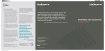
HallmarqStanding Equine MRI HallmarqStanding Equine MRI Are all MRI scanners the same? There are three types of MRI scanner: • Standing MRI. The Hallmarq system is the only MRI scanner that can image the foot and leg of the standing sedated horse. Almost all equine MRI systems in the UK are Hallmarq standing units. • Adapted human 1.5T high field, tubular scanners. Mostly found in institutions that scan both companion animals and horses, there are very few of these in the UK or Europe. The reported diagnostic rate and lesions detected are similar for both high field and standing systems, though some clinicians prefer a high field machine for performance issues where only minor lesions are suspected. General anaesthesia is always required. • Low field "down" scanners. General anaesthesia is again required, though without gaining the benefit of the stronger magnetic field. Some such systems can scan body parts larger than the distal limb. Is MRI safe? There is a known* risk that about 1 in 200 otherwise healthy horses will die or suffer complications due to general anaesthesia or on recovery. Horse owners are aware of the risk and often reluctant to consider general anaesthesia for a diagnostic procedure alone. The Hallmarq MRI system was specifically designed to be safe, with no need to anaesthetise the horse, lay it down, or move it using hoists. *1. Johnston, G. M., Taylor, P. M., Holmes, M. A. & Wood, J. L. Confidential enquiry of perioperative equine fatalities (CEPEF-1): preliminary results. Equine Veterinary Journal 27, 193-200 (1995). Is MRI expensive? On average standing MRI proves no more expensive than conventional diagnosis. Lameness is a challenging condition, and often a horse undergoing just conventional work-up and treatment will return to the clinic again and again, accumulating higher total costs than an early, definitive MRI diagnosis. Early diagnosis is also likely to leave funds available for more effective treatment, improve outcome, and reduce distress to horse and owner. How do you deal with motion? During a standing foot scan the foot is placed firmly on the floor, and with correct positioning the horse will usually remain remarkably stationary for the 2-5 minutes required for each set of images. Any unsatisfactory scans can quickly be identified and repeated. Higher up the leg Hallmarq's awardwinning motion correction software compensates for movement, and repeats any parts of the scan that would lead to blurring. "Whilst certain clinical, radiological and ultrasonographic findings may point toward a particular diagnosis of foot pain, in many horses with foot pain an accurate definitive diagnosis can only be made using MRI1 Parkes R., Newton R., and Dyson S.J. Vet J 204, 40-46 (2015) To take a look at some interesting case studies please visit: www.hallmarq.net/equine/cases-studies "Standing MRI allows us to accurately diagnose the cause of lameness in the vast majority of cases where standard diagnostic techniques fail to give us the answer. It permits the selection of appropriate treatment methods, whereas without it we would often have been guessing" Tim Mair BVSc PhD DEIM DESTS DipECEIM MRCVS, Bell Equine Veterinary Hospital REFERRING FOR EQUINE MRI Standing for Safety Hallmarq Veterinary Imaging Ltd Unit 5, Bridge Park, Guildford, Surrey, GU4 7BF, UK +44 (0)1483 877812 Hallmarq Veterinary Imaging, Inc. 1275 W. Roosevelt Rd., Suite 116 West Chicago, IL 60185, USA +1 978.266.1219 hallmarq.net | [email protected] mr © Hallmarq Veterinary Imaging 2018.
Open the catalog to page 1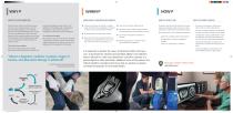
WHAT'S SO SPECIAL ABOUT MRI? WHEN SHOULD I CONSIDER AN MRI REFERRAL? Acute onset of lameness during exercise Racehorses at risk of fetlock fractures or palmar osteochondral disease through repetitive fast work Cases that do not respond to treatment as expected Monitoring the progress of treatment, and to assess readiness for competition Since the advent of MRI, much has been learned about the causes of equine lameness. From the previously under-diagnosed, such as collateral desmitis of the distal interphalangeal joint, through the previously misunderstood, such as navicular syndrome, to the previously...
Open the catalog to page 2All Hallmarq Veterinary Imaging catalogs and technical brochures
 2018 STANDING EQUINE MRI
8 Pages
 Veterinary CT
3 Pages
 SMALL ANIMAL MRI
5 Pages
 Escala saher culivators
2 Pages
 MIST BLOWERS
4 Pages
 ATOMIZERS
4 Pages
 SPRAYERS
4 Pages
 2016 STANDING EQUINE MRI
8 Pages


